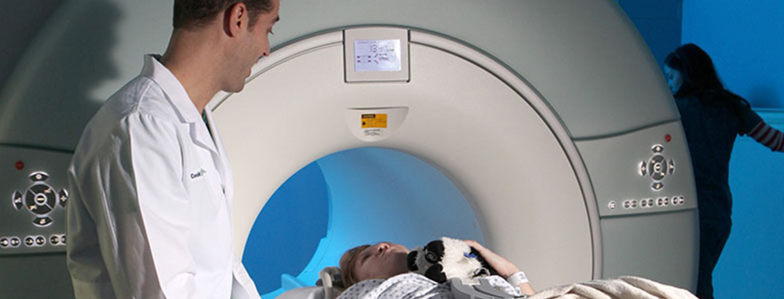
The MRI is forty-seven year old. The concept of MRI was first conceived by Isidor Rabi, a physics professor at Columbia University in 1937. Rabi developed a method for measuring the movements of atomic nuclei – a state he decided to call nuclear magnetic resonance (NMR) and was awarded the 1944 Nobel Prize in Physics. Rabi’s method was used only for chemicals.
The image-producing scan we know today was developed in 1973. MRI was originally called NMR. The first concept of Magnetic Resonance Imaging was called Nuclear Magnetic Resonance, or NMR. The name was changed because of the negative connotation of the word “nuclear”. MRI machines are calibrated in Tesla units to honour Nikola Tesla, who discovered the rotating magnetic field in 1882.
MRI can diagnose small fractures. MRI scans are often used to identify tumours or bone fractures that are too small for an x-ray. These are often called hairline fractures. Since an MRI does not use radiation technology, they are safe enough to use for research purposes. They are commonly used in clinical research to study the brain activity of individuals performing day-to-day activities. This research study is called fMRI. Functional MRI (fMRI) evaluates brain activity by detecting changes linked with the blood flow. This technique depends on the fact that cerebral blood flow and neuronal activation are combined. When an area in the brain is utilized, blood flow to that region also increases.
Kryptonite has introduced this latest technology to the range of MRI products. It is a turnkey fMRI Solution with user-friendly interface & fMRI protocols as per ASFNR standards. The use of fibre optic technology for the delivery of the signal to the shielded screen enclosure guarantees an artefact-free scan. As the system is permanently mounted there is no setup required when functional imaging is taking place ensuring an efficient throughput of patients. The Stimulus software provides the ability to customize paradigms as required. The patient response system helps in getting feedback with real-time synchronization with the MRI scanner.
Many people are averse to the noise and confined space of an MRI machine, which is typically an enclosed tube. The creation of In-Bore MRI Cinema has made the experience more pleasant for children and adults those who suffer symptoms of claustrophobia, and enables doctors and family members to talk and see the patients while the scans are carried out. In-Bore MRI Cinema is mainly designed to lower patient claustrophobia & improve their MRI experience. The atmosphere created by In-Bore MRI Cinema diverts the patient’s attention giving a very soothing experience to the patients. Also, it gives your patients the freedom to relax & stay calm inside an MRI scanner with high-quality entertainment of their choice.
Patient comforts through sound and Kryptonite’s MRI Audio system is a state of a art audio system that helps reduce anxiety and claustrophobia during noisy and stressful MRI scan procedures. Patients can relax and listen to their favourite music, listen to videos through noise cancelling headphones. Easy to use for technologists, the system also allows direct communication with patient at any time during scan. It also reduces the need for re-scans, call backs or sedation medicine.
The MRI Audio system includes everything imaging centres need to relax nervous MRI patients before and during scans. The system can be easily customized and fully upgraded by any imaging centre at any time, future-proofing it against the rapidly changing landscape of audio entertainment. As a complete audio system, it’s an effective means of enhancing patient enjoyment while improving satisfaction scores. This leads to more efficiency and profitability for the MRI centres.
MRI, being of the most common imaging scans, presents a challenge for many patients. MRI Ambient Experience for MRI solution already contributes to a positive patient and staff experience. Patient experience is an important factor for the successful operation of a radiology department. Patients are positive and the MRI staff see these features help calm patients, reducing motion-related problems and providing excellent images. These are some of the tangible results which can help your healthcare facility achieve with Ambient Experience. It demonstrates that reducing patient stress can make it easier for them to cooperate and thereby decrease delays and retakes. This can all have a remarkable impact on your clinical, operational and financial challenges.
MRI Ambient Experience was proposed to transform the traditional scanning experience, it gives patients a comforting and engaging experience during the scan. The focus on patient experience has helped the Department of Radiology in many Hospitals to differentiate itself from others in the region. Patient healing and department efficiency go together.
Evidence based research approach and people-centric design thinking have helped facilities uncover new opportunities to improve the patient, family and staff experience. MRI Ambient Experience can benefit many areas from waiting areas and uptake rooms to treatment and recovery rooms and even entire departments. Radiology department uses room design products like Ambient Lighting and MRI Projector to give patients greater control and positive distractions during their journey.
The most desirable feature in any architecture is a visual connection to nature. When building design or location interdicts this life-supporting feature, a biophilic illusion of nature restores this essential wellness benefit. It is our contention that the properties of illusory images generate a deeper relaxation response in the observer than has been documented employing standard representational imagery.
We also contend that such illusions can change the subjective experiences of space in interior environments. The only evidence of this psycho-physiological phenomenon was based on the testimony of patients and others who reported experiencing a palpable feeling of openness and expansion that went beyond what is accomplished with decorative nature imagery in healthcare.
Session delays or cancellations, in addition to MRI retakes due to the patient’s trepidation and anxiety about the procedure, raise a hospital’s average cost per session. Since imaging sessions are among the far most expensive environments to operate, environmental design features that can reduce patient’s stress and help them relax and remain still, will improve an operator’s MRI efficiency.
MRI magnets get very hot, so the magnets used in MRI must be cooled. Liquid nitrogen is used to take the magnets down to 0 degrees Fahrenheit. MRI magnets are strong. The main magnet in an MRI magnetic field is 140,000 times stronger than the Earth’s magnetic field.
Most people have no idea what imaging analysts’ duties consist of Post-Processing and Image Analysis. The job is actually pretty elaborate. Employees in the Radiology department must have training in Radiologic medical imaging. Employees in the radiology department must exhibit proficiency in the utilization of electronic tools, paying close attention to details while creating and preparing imaging manipulation on virtually every part of the body. They work on a variety of software programs, acquiring extensive knowledge of human anatomy.
All imaging analysts previously worked or continue to work as MRI technologists should maintain certification in MRI modalities. MR images can be manipulated for evaluation in various ways like MRI Post-Processing. Post-Processing of MRI includes: Subtraction, addition, rotation, inversion, multiplanar reconstruction (MPR), maximum intensity projection (MIP), etc. Olea Medical is providing the most advanced post-processing solutions for MRI. Together, they offer the best of both worlds.
Olea is one of the leading companies in developing clinical applications for advanced post-processing of MRI, Olea Medical’s innovative solutions allow for image viewing, analysis and processing of even the most complex MRI sequences, and standardizes both viewing and analysis capabilities of functional and dynamic imaging datasets acquired with MRI.
Moreover, the new Olea Vision developed by Canon Medical and Olea provides the basis for more than 40 completely new and unique applications. All applications can be viewed by using Olea Medical’s viewer Olea Vision. As these automated systems become pervasive in the healthcare industry, they may bring about radical changes in the way radiologists, clinicians, and even patients use imaging technology to monitor treatment and improve outcomes.
If there are some kinds of metal objects inside your body, the MRI’s magnetic field may be a problem. The MRI technician will ask you regarding all metal objects that are inside your body. Some metals are safe and some are not. The technicians have a detailed list of what’s safe for a given MRI machine.
But in general, MRI is a problem for Medical devices controlled by magnets, such as a heart pacemaker, defibrillator, or cochlear implant – the MRI can make the device malfunction, Medical devices with wires or other metals that conduct electricity – the MRI can make the device heat up and burn you and Metal, such as iron, that can be pulled by a magnet – the MRI can make this metal pushed inside your body.
Kryptonite’s Magnetic Resonance Imaging (MRI) compatible products are used throughout the MRI suite to help facilitate MRI use. Our MRI safe products help maximize safety and comfort—for both patients and medical staff. Improve patient care with top-quality products from MRI transport equipment to MRI furniture, Immobilizers, Pulse Oximeters, Fire extinguishers, MRI Phantoms and more.
Since the three major commercial MRI vendors (General Electric, Philips and Siemens) started to produce ultra-high field (UHF) MRI scanners for human imaging in circa 2002, the installed-base has increased rapidly, reaching approximately 55 sites with systems of 7T or above. This rapid growth in the number of UHF-MRI sites has quickly led to the development of scanning protocols and the required hardware to enable the use of UHF-MRI in both fundamental and clinical research studies as well as in pilot studies for pure clinical use.
Read more – What is fMRI
MRI audio systems let patients listen to music or calming sounds during their scan, helping reduce anxiety and make the experience more comfortable.
They’re made from non-magnetic materials and use special cables or wireless tech to safely deliver sound inside the MRI room.
Yes, many systems let you pick your favorite music or even stream from your device, making the scan more enjoyable.
Absolutely! Listening to music or calming sounds can distract you from the noise and help reduce feelings of claustrophobia.
Yes, they’re designed to fit all ages and are safe for children, helping make their MRI experience less scary.
Yes, most systems have a feature that lets the technician communicate with you clearly, even while you’re listening to music.
They help by keeping patients relaxed and still, which leads to fewer motion artifacts and better scan results.
Besides audio systems, there are advanced lighting, themed ambience, and interactive displays to further improve patient comfort and experience.
©2024 Kryptonite SolutionsTM. All Rights Reserved.
Powered by: Purple Tuché