Brain imaging allows doctors and scientists to see the inner structures of the brain without having to open the skull. There are several brain imaging techniques. One is called magnetic resonance imaging (MRI), which examines the structure of the brain, and another is functional magnetic resonance imaging (fMRI), which examines brain function. Functional MRI measures brain activity by tracking changes in blood flow to the brain.
Functional MRI is derived from structural MRI. They both use the same device called an MRI scanner. MRI technology is used to create detailed 3D images of an object’s internal structure using magnetic fields and radio waves. MRI can be used to examine body parts other than the brain and even non-living objects. Brain MRI and fMRI are used in medicine to diagnose diseases, plan treatments, and study the underlying causes of diseases and disorders.

How does an MRI scan work?
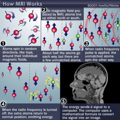
MRI scanners work by photographing a thin slice of the brain at a time. The images are then stacked like pancakes to create a complete picture of the particular region. The human body is made up of billions of molecules, including water molecules, MRI-compatible monitor machines can detect that. The atoms in all molecules, including water (H2O) molecules, contain protons. Protons are like little magnets. In the absence of a very strong magnetic field (i.e., when we are outside the MR scanner), the protons in our body orient themselves in random directions. When we are inside the scanner, its powerful magnetic field, typically ten thousand times stronger than Earth’s magnetic field, forces these protons to align with the field, although we cannot feel it at all.
The gradient coil helps scanner operators pinpoint where our body is within the scanner. Then the radio frequency (RF) coils emit radio frequency waves in all areas of the imaged body to realign these protons again, but temporarily. The RF coils can be part of the MRI machine for full body scans or used as a special head coil when only imaging the brain. When radio frequencies are no longer transmitted, the protons “relax” to their original alignment with the scanner’s magnetic field. As they do so, the protons release the energy that pulled them toward the RF coil in the form of electromagnetic signals.
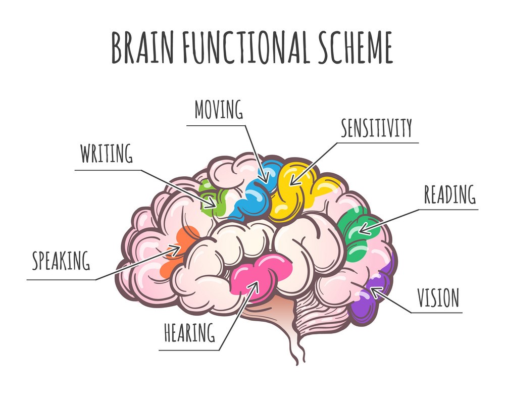
While MRI only takes pictures of brain structure, fMRI shows brain activity (or function) by comparing blood flow under different conditions. It allows us to interpret the things we see, touch, hear and taste and regulates our body’s reactions to the external environment. All of this happens through networks of tiny cells called neurons, which process and transmit information between the brain and the rest of the body. When the brain is presented with a task, such as remembering an idea, the neurons responsible for that activity become more active than other neurons around them. They generate chemical and electrical signals and transmit them from one neuron to another. This process is called neural activity or brain activity.

How does Functional MRI measure Brain activity?
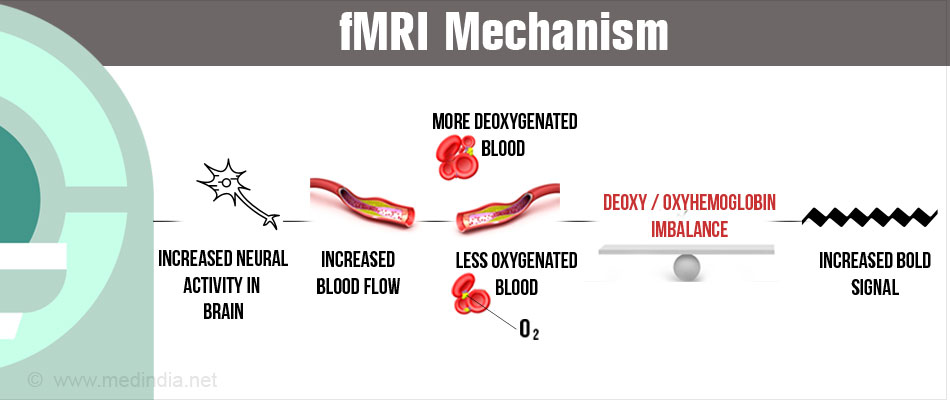
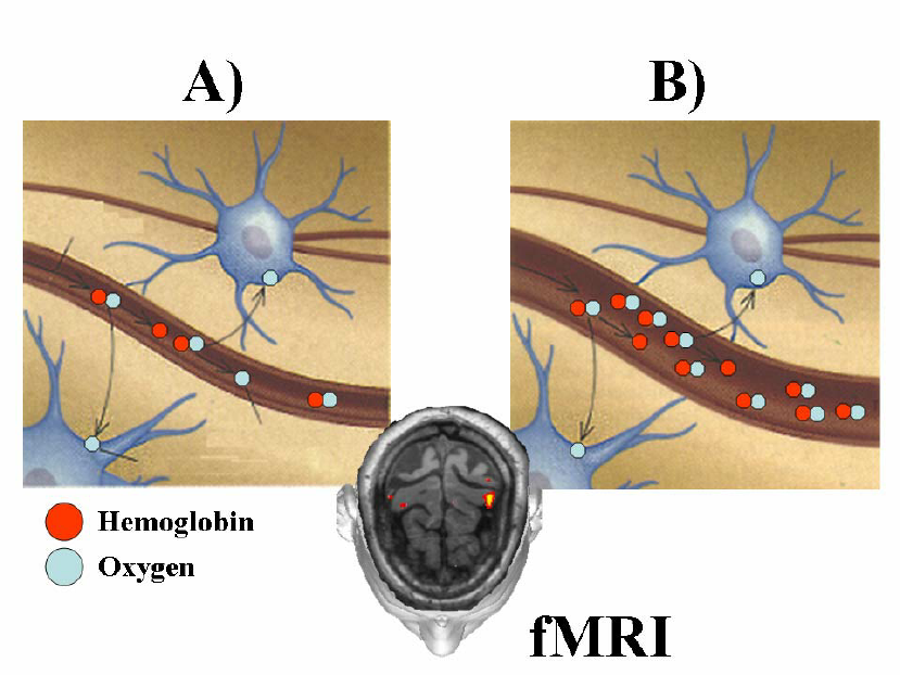
Functional magnetic resonance imaging, or fMRI monitor, is perhaps the best-known technology for recording neuronal activity, but it doesn’t actually record the activity of neurons; instead, the multi-coloured images you see of certain brain regions that light up reflect blood flow in the brain. More specifically, the signal you see reflects the relative presence of oxygenated versus deoxygenated blood; Active regions require more oxygenated blood, and thus fMRI, although indirect, allows scientists to infer patterns of neuron activity. and function correlates in humans.

More energy is used when neural activity increases in one part of the brain. More oxygen-carrying blood is needed to replenish this energy. Transported to this brain region. Blood carries oxygen via a molecule called haemoglobin. Haemoglobin contains iron, which gives it magnetic properties, like a small iron filing. Depending on whether haemoglobin is oxygenated or not (i.e., whether it is oxygenated or deoxidised), it has slightly different magnetic properties. Therefore, increased neural activity occurs. With a greater flow of oxygenated blood, so the more active regions of the brain are slightly more magnetic. 
Functional MRI detects brain activity by measuring changes in the amount of oxygen in the blood and blood flow . This measurement is known as blood oxygen-dependent activity (BOLD activity). In other words, BOLD activity is a convenient surrogate for brain activity: fMRI visual system indirectly measures brain activity by measuring BOLD activity.
Applications:
The development of MRI and fMRI has enabled us to make great strides in understanding brain function. One of the reasons for this is that MRI and fMRI, unlike X-rays, for example, do not damage brain cells with harmful radiation and can pinpoint the location of brain activity more precisely.

For example, just a few years ago, we could only track Alzheimer’s disease at later stages, when most of the damage to the brain is irreversible. People with Alzheimer’s tend to have less activity in certain parts of their brain compared to healthy people. Therefore, doctors can use fMRI to diagnose Alzheimer’s early by detecting abnormal or lower activity levels in these brain regions before the disease worsens. In addition, fMRI scans can now distinguish between a brain with and without autism with an accuracy of 97%.
fMRI studies have revealed patterns of decreased activity in the part of the brain important for planning, problem-solving, and interpreting social interactions. In addition, scans of autism patients have shown fewer connections between the brain’s two hemispheres. Therefore, rapid advances in understanding these diseases will be made by systematically comparing brain function in patients with certain brain-related diseases to brain function in healthy patients.
Challenges:
Nevertheless, fMRI lets in an unrivalled a take a observe wherein and to what quantity distinct capabilities can be localised in the human brain, and researchers retain to plot approaches to enhance its spatial and temporal resolution, as an example via way of means of making the method touchy to neuronal adjustments in preference to adjustments in blood flow. No contemporary approach suits fMRI for its cap potential to ‘map’ or decides the probably supply of cognitive features in the human brain. Despite all of the progress, some demanding situations remain. For instance, we nonetheless recognise little or no approximately how the mind develops in early childhood.
Currently, fMRI machines are dark, noisy, and scary, making them mistaken for toddlers and those frightened of cramped spaces. They additionally take too lengthy to create snapshots, and their “cameras” are extraordinarily touchy to movement, making the scanned images blurry. Furthermore, fMRI is expensive, now no longer portable, and calls for a variety of education for medical doctors and scientists to use. Researchers are running to resolve those problems. Kryptonite Solutions
©2024 Kryptonite SolutionsTM. All Rights Reserved.
Powered by: Purple Tuché