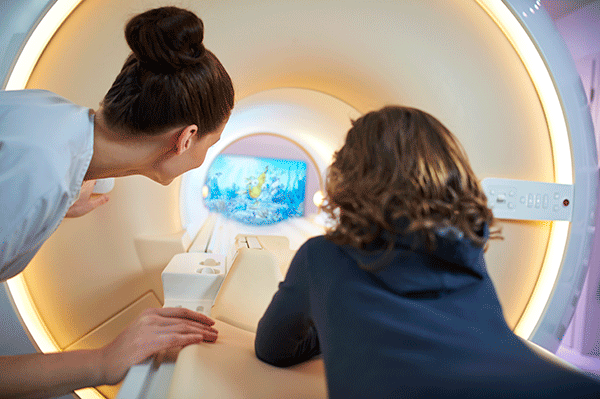
Magnetic resonance imaging expertise involves plenty of various areas that might be polished to improve the patient experience. Below follows an inventory of things that are a part of the experience:
All of the above have been modified throughout the development of magnetic resonance imaging to boost the patient experience. Some areas aren’t as vital as different and have thus not been targeted neither within the development of the product on the market nor in the scientifical field. Such areas are, for example, lying in a very bound position. Though the bore that the patient lies on within the MRI has been changed to be ready to lie additionally still, since the primary MRI till today, there are other areas wherever more has happened. 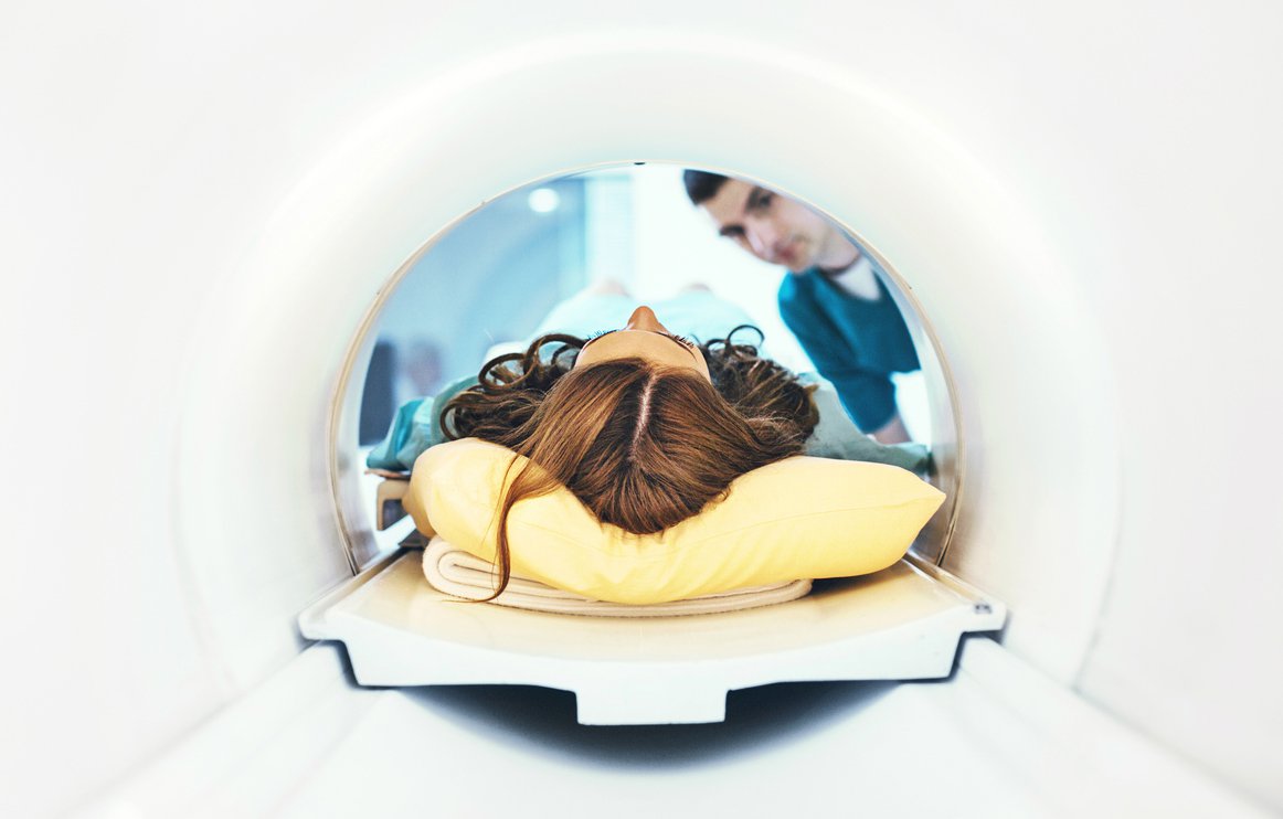 MRI narrow bore
The narrow hollow tube: The patient must lie in a very narrow tube throughout the scan. When patients suffer from claustrophobia, which many suffer from, the narrow tube is a trigger for anxiety. Solutions have been found to reduce this impact, therefore, creating a short hole that is wider and tapered in shape. This should reduce the claustrophobia that occurs. Since a wider tube poses difficulties in generating a homogeneous magnetic field, the tube’s width still has limitations. 
Long scan time
The Scan Time: The examination time of an MRI is considerably longer than an X-ray and CT examination. For comparison: It only takes a minute to create a CT image. CT scan times vary depending on the target but can be as short as ten minutes, while a fast MRI takes about 20 minutes. A study shows how anxiety levels change over time. This study measured anxiety by monitoring patients’ heart rates, with higher heart rates indicating higher stress and anxiety levels. The result showed that the heart rate was highest during the dissection and when the hole was moved to the scanner, not during the scan itself.
The longer anxious patients were in the scan, the heart rate decreased and stayed constant at a low level throughout the scan for non-anxious patients. Then, after the scan, the anxiety level rose again, although not to the same level as before the scan, before returning to normal. The result of the heart rate test was supplemented with the patient’s subjective anxiety. The conclusion was that a solution is needed to reduce anxiety before and during the transition to MRI. 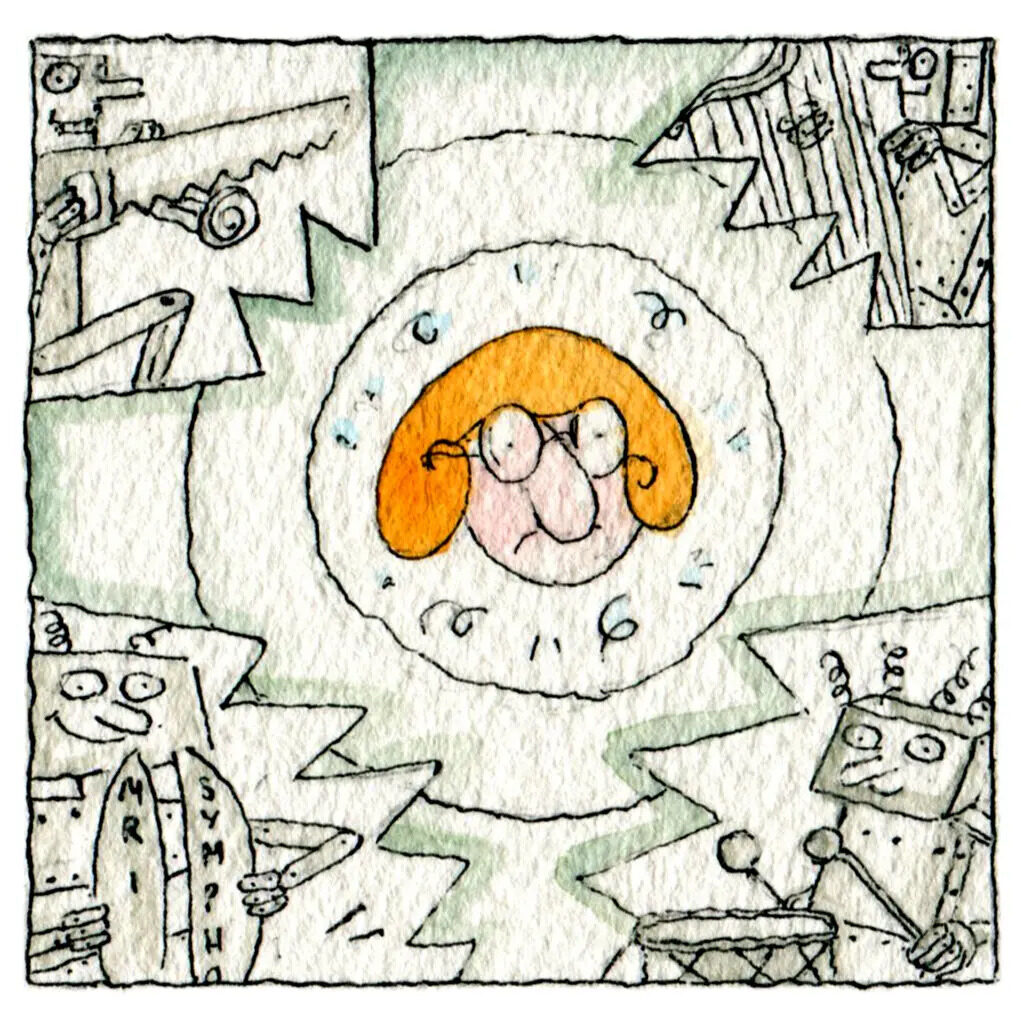

The noise: A conventional MRI emits noise of up to 128dB. In context: If you are standing in front of a rock concert, your sound exposure is 120 dB. 120 dB is also the ear pain threshold. Not only is noise uncomfortable and harmful, it also increases the incidence of claustrophobia. Therefore, noise reduction is important if the patient experience is to be improved. Studies show that noise reduction positively affects the image even when the patient is sedated. There are different methods to achieve the same goal: Use noise cancellation headphones or reduce the volume of the MRI machine. Although there are published articles on recently developed MRIs with noise reduced to 77 dB, due to industry involvement in this research.
There are several studies on how the headphones used in MRI could be improved. A normal noise-cancellation headset could not be used due to the magnetic field in the MRI. It is also worth noting that this reduction is measured and compared within the earpiece, so the overall sound reduction will be a combination of this active reduction and the passive reduction of the headphones. This reduces the weight of the headset and improves the patient experience. Since the ANC technology is in a headset, this can be done without investing in a new machine. Instead, the ANC technique can be used in any existing MRI setup. 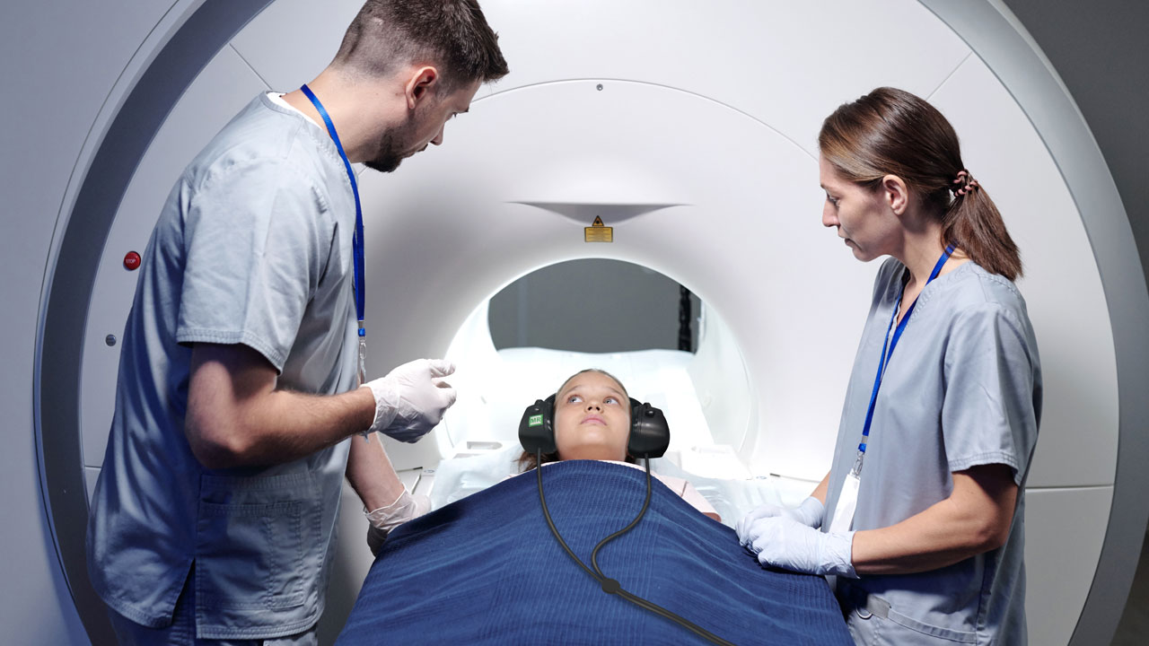 MRI audio system: Thus, this noise cancellation technique can reduce the noise and improve the patient experience by implementing the MRI- compatible audio system.
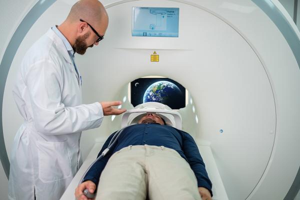
The focus on the patient’s vision: Patients cannot look around during the scan because they have to lie still. The only thing the patient can see is the roof of the narrow tube. Even with relaxing sounds, focusing on anything other than the MRI is difficult as that is all the patient can see. 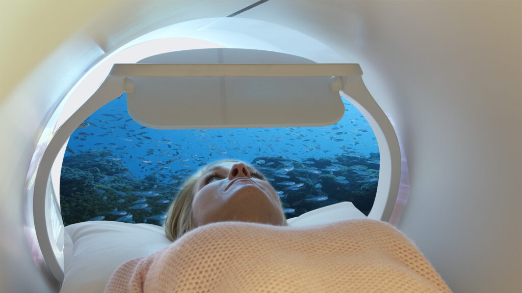
Vision is one of the core senses during MRI, and there are ways to improve it, but little to no scientific research has been found in this area. However, there are several solutions on the market using MRI-compatible goggles or more like a helmet with an angled mirror placed in front of the patient’s eyes, allowing the patient to look through the tube in a supine position. By placing the MRI-compatible monitor behind the bore, the patients visualize the monitor screen through the mirrors or goggles. This brings a whole new patient experience of watching a TV inside an MRI. The patient can view anything of their choice while the scan is done. This MRI In-Bore Experience reduces patients’ anxiety and claustrophobia, thereby reducing the scan time and rescans. 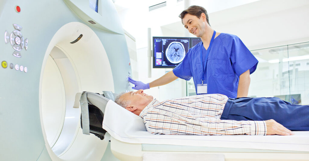
Communication with the patient: Many patients fear the procedure because they do not know what will happen. Therefore, communication with the patient is important, both before the examination to prepare for it and during the examination to reassure the patient and let him know how he is doing. There are many different solutions to this problem. Starting with informing the patient earlier, one study shows the effect of sending the patient DVDs at least a week earlier. The DVD was in addition to the standard care and included two clips. a bracket was a seven-minute prep video, and the other clip included 12-minute relaxation exercises. Information and the patient’s ability to self-manage, when stressed during the scan, could be related to lower amounts of motion artifacts compared to the baseline group. 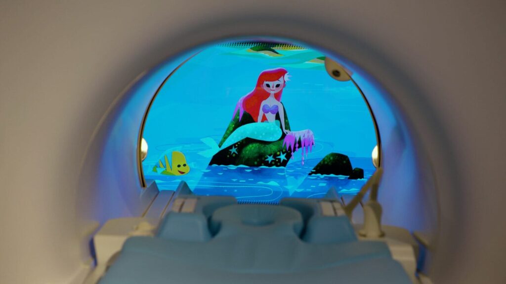
Entertainment for children: It has also proven useful for children to show some kind of film before scanning. It examined how a three-minute animated informational film, the ideal length for educational videos, affects children before scanning. The footage was found to help children understand and prepare for MRI. This is not just something that comes up in reports, vendors have already developed solutions to these insights.
Several studies also write about the importance of information during the scan. The MRI scan consists of several sequences; between these sequences, the patient can communicate with the staff as long as they remain silent. This can be done via an intercom with a microphone in the technical room where the technologists are located and one in the MRI room. They indicated that the group that could communicate every two minutes between sessions showed less anxiety.
The MRI environment: All the pieces come together to create a relaxing atmosphere for the patient. It all starts when the patient enters the MRI room. The importance of a relaxing sound has already been mentioned. Another thing that has been proven to enhance the experience is the lighting in the room. Lighting in hospital environments is often a complex matter. Patients need a light that calms and promotes the healing process. During an MRI, the doctor doesn’t need bright light to see, so the focus may be on calming the patient. This can be done with a variation of Patient relaxing lights called a virtual skylight. Odours in the room can also affect the atmosphere and thus the patient experience.
©2024 Kryptonite SolutionsTM. All Rights Reserved.
Powered by: Purple Tuché