☰

INTRODUCTION: The MRI Safety Committee established the MRI SAFETY Guidelines to imply safety policies for all research and clinical MRI systems. The Safety procedures are carried out every month by the MRI Safety Committee. The MRI Safety Guidelines are intended to provide information on safe Clinical use and Research of MRI systems. It also assists in practising the appropriate protocols and procedures of MRI. Therefore, it also helps in preventing adverse patient outcomes related to MRI. The guidelines help the Doctors, Radiologists, Radiographers, Fellow members and Nurses working in the MRI department address the safety issues facing the MRI department. The main goal of these guidelines is to provide the accurate, proper, and safe use of Radiological services for the betterment of patients by leading and training our Doctors and Radiation Professionals.
MRI Safety Officer: The MRI Safety Officer is a person responsible for the formulation and application of policies and for assessing the risk and benefits of unusual situations. The safety officer also ensures the safety of the patients, radiological staff and others working in the department. The MRI Safety Committee appoints an MRI Safety Officer for MRI safety-related tasks and to imply its site`s safety policies. The safety officer must be trained in the MRI, and MRI Safety and must be well experienced in the MRI environment.  MRI Safety Education: The MRI Safety Officer should conduct annual training and presentation to all the staff working in the MRI environment. The officer must conduct routinely training for the doctors and technicians working in zone 3 and zone 4 based on the following topics:
There should be annual training given to the Hospital staff working in the other department who interact with the MRI department on the MRI basic safety principles and MRI zones. 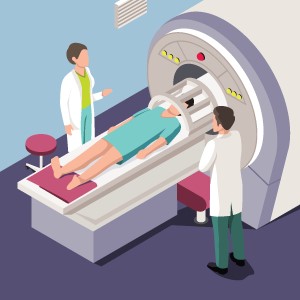 MRI Environment: A policy is implemented by the MRI Safety Committee to ensure the restriction of access to MRI Scanning and control areas. This policy aims to the use the following zones and the restrictions to help serve the following requirements:
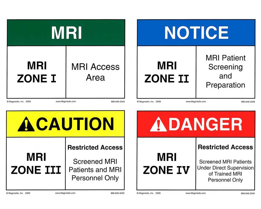 FOUR-ZONE SYSTEM:
Zone 1: There are No Restrictions. It is the general public area outside the MRI department. This area is the reception and the patient waiting areas.
Zone 2: It is the area between Zone 1 and the strictly controlled Zone 3. The safety screening usually occurs here. The zone 2 is that area of travel where patients are brought into their procedure and the interception and safe storage of all removable ferromagnetic or potentially ferromagnetic objects should take place in this area.
Zone 3: It is called as the Control Room. All the access to the control room is strictly prohibited with access to regions controlled by the MRI Personnel or Radiographers. This zone 3 is strictly restricted to the general public and will differentiate the MR and Non-MRI professionals. 
Zone 4: It is the MRI Magnet Room. No one is allowed inside the MRI scan room without being instructed by the MRI Professional or Radiographer. The patient and its relative can enter the scan room during the scan by thoroughly supervised by the MRI Radiographer. The MRI machine room door is always locked and at times unlocked the MRI Safety device like Safety Barrier must be implemented by the MRI personnel. The MRI compatible equipment can only be brought in the MRI scan room under the guidelines by the MRI Safety committee. In an MRI department, all the patient transfer activities must run through these 4 zones indicated above.
The patient transfer should occur in the Zone 2 that is the Holding area. In case if there is no holding area, the patient transfer must occur in Zone 3. Before taking the patient in the scan room for the MRI, a procedure is carried out in following steps:
 Potential Hazards and Risks:
In such a Strong magnetic field, objects that are prone to attracting the magnets such as Small clips, hairpins also pose a potential risk to the patient inside the MRI room. Sometimes, even surgical tools such as Scissors, Haemostats, and clamps that are in the MRI department made of stainless steel attract to the Magnet. Objects such as Oxygen cylinders, Floor cleaning equipment, patient trolleys and other construction tools which are strongly attracted by the magnet should not be brought into MRI department.

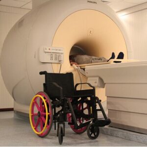
To avoid such risks, MRI Safe Equipment made from non-ferrous materials such as oxygen cylinders, patient trolleys, wheelchairs are MRI compatible.
 Magnetic Quench: Quenching is the process where there is loss of temperature to zero in the magnetic coils, thus eliminating the magnetic fields. This leads to the escape of Helium gas from the Cryogen bath in rapid pace and resulting in entering the MRI scan room. Quench leads to the depletion of oxygen in the MRI room, thereby suffocating the patient and sometimes to death. It may happen in accidental conditions or done manually in case of Emergency.
Acoustic Noise: Acoustic Noise is generated when current runs through the gradient coils during the MRI scan. The noise generated by the gradient coils has noise levels below the OSHA standards for a hearing loss prevention programme must be started. The noise can have effects such as patient annoyance, hearing loss, hearing impairment and sometimes permanent loss. All the patients are given earplugs, or headphones
MRI Contrast Administration Policy: The administration of Contrast depends on the radiologist by assessing the condition of the patient, and whether the contrast is essential or not. The contrast administration is determined by reviewing the initial findings in non-contrast images. A trained professional must administer the contrast agent to the patient.
Patients who are at increased risk of Contrast:
Precautions to be taken by the Kidney disease patients: Patients with impaired kidney function, the MRI scan without the Gadolinium based contrast, or with another modality should be considered. In order to carry out the MRI scan with contrast agent certain precautions are taken in to consideration:
Retention of Contrast (Gadolinium): It is noted that in some cases, after the contrast administration in MRI, there is mall amount of contrast agent like gadolinium is retained in the body, even in normal kidney function patient. Such cases of Gadolinium occur to a great extent with high-risk agents causing the abnormalities in certain part of the brain. However, it is safe to administer gadolinium only when it is reasonably expected to give some additional meaningful clinical information. 
MRI in Pregnancy and for lactating mothers: With the increase in the use of MRI scan for the clinical information of patients, results in an increase in requests for MRI scans in Pregnancy and lactating. The main objective of the MRI Safety Guidelines are as follows:
In cases of Pregnancy and Lactation, there is a risk of undergoing an MRI scan due to exposure to electromagnetic waves.
For Pregnant patients: In pregnant patients, before undergoing any scan consent is taken by informing the patient about the scan and its risks. In the first trimester of Pregnancy, the MRI scan is done for maternal rather than the foetal indications, and in such situations, MRI is preferred as compared to CT scan. In contrast study of the MRI during pregnancy, there is very less evidence of the safety of contrast in pregnant patients. The injected contrast reaches the amniotic fluid, where its presence and its effects are unknown.
For Pregnant medical staff: Working in and around the MRI environment during pregnancy for medical practitioners is permitted. Some activities are accepted but limited to the control room in the MRI. Pregnant workers are permitted not to work within the MRI bore or scan room during the data acquisition or scanning.
For Lactating Mothers: As Safety precautions, the gadolinium-based contrast is avoided, but excretion of the contrast into breast milk is shown to be minimum. Therefore, contrast for lactating mothers is not absolutely contraindicated, and breast milk need not necessarily be discarded after contrast administration.
©2024 Kryptonite SolutionsTM. All Rights Reserved.
Powered by: Purple Tuché

