An MRI is a painless, non-invasive and a safe medical procedure. An MRI or fMRI is a fast, efficient and highly detailed, and a very valuable diagnostic tool that can help the doctor accurately assess and effectively treat a wide range of medical conditions.
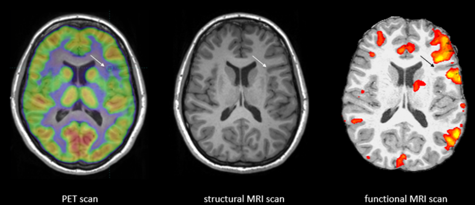
Functional MRI is a part of Magnetic Resonance Imaging that deals with the brain`s functional activity in the rest and the active states. fMRI (functional Magnetic Resonance Imaging) is a method used for brain activities and injuries such as concussions than a standard MRI (Magnetic Resonance Imaging) brain scan.

Magnetic resonance imaging (MRI) is a type of medical imaging that uses strong magnetic fields to produce images of the body organs. MRIs are used to assess a variety of conditions in many parts of the body. For example, an MRI of the brain and spinal cord can reveal evidence of: Brain injury, Stroke, Blood vessel damage, Cancer, Multiple sclerosis, Spinal cord injuries. A scan of the heart and blood vessels may show: Heart disease, Blocked blood vessels, Abnormal heart structure. Bones and joints can also be imaged using MRI technology, detecting: Cancer, Joint disorders, Problems with the intervertebral discs, Bone infections. An MRI can also be used to evaluate organs such as the liver, kidneys, breasts, pancreas, pelvis, and prostate.
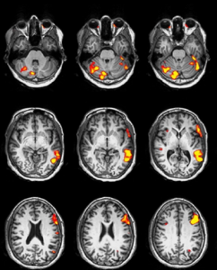
Functional magnetic resonance imaging is a specialized form of MRI that is used for measuring and mapping the brain’s functional activity, meaning which part of the brain is handling critical functions such as speech, motor, listening, etc. An fMRI measures changes in blood flow that occurs in the brain to detect areas of activity. The primary reason for an fMRI scan is to help the doctor identify the activated regions of the patient`s brain before they go into brain surgery. The scan will help doctors better understand the regions of a stroke or other condition affecting the brain or spinal cord.
The fMRI measures the changes in those electromagnetic waves, so if blood flow levels change in a particular region (signalling increased activity), the fMRI will show the change (known as a BOLD signal). We can then translate this into a brain map of the observed tissue that shows where blood flow, and thus brain activity, is and is not happening when a patient performs a task. In contrast, a structural MRI provides information about brain anatomy to complement fMRI. The anatomical information provided by the structural MRI acts as a reference for the visualization of activated regions of interest. Combining the information from a structural MRI and fMRI may broadly characterize normal and abnormal brain functions.
We would like to introduce to our latest technology of fMRI products. It is the state-of-the-art Turnkey fMRI solution with the protocols based on the ASFNR standards with the user-friendly interface and custom-built experiments. As our In-bore system can be permanently mounted on the wall there is no additional setup required every time when the functional scan is performed thereby increasing the efficient patient work flow. Our In-bore Monitor system can be connected with an HDMI output of any computer system.
To deliver the signal to the shielded screen we use the fibre optic cables whose enclosure guarantees an artefact-free scan. The patient response feedback system helps with the real-time synchronization with the MRI scanner. We also provide with the state-of-the-art fMRI post processing solution so that the recorded data yields the functional information which is useful for the doctors. Our fMRI System Package consists of:
Visual presentation is displayed on an MRI compatible visual display system placed in the exam room, with sound routed into your existing scanner headphones via the technologist intercom system. Simply, play the experiments and the patient has to perform the activity displayed on the Visual system.
The Stimulus presentation software runs the experiments installed as per ASFNR standard protocols which the patient has to perform. It supports audio and visual stimuli creation and delivery, and records responses from nearly any input device. Patient Response Pads The fMRI response pads are used by patients to respond to the experiments or activity presented by the stimulus software. It is provided with multiple buttons options to ease the patient`s response activity.
Synchronization Box is able to condition and distribute a SYNC signal (analog and digital) to different devices simultaneously, from the MRI scanners to the stimulus software and the response pads.
Brain activations can be generated from fMRI raw data sets by applying several mathematical methods. The post processing application works as a tool for the analysis of anatomical and functional MRI data sets.
©2024 Kryptonite SolutionsTM. All Rights Reserved.
Powered by: Purple Tuché