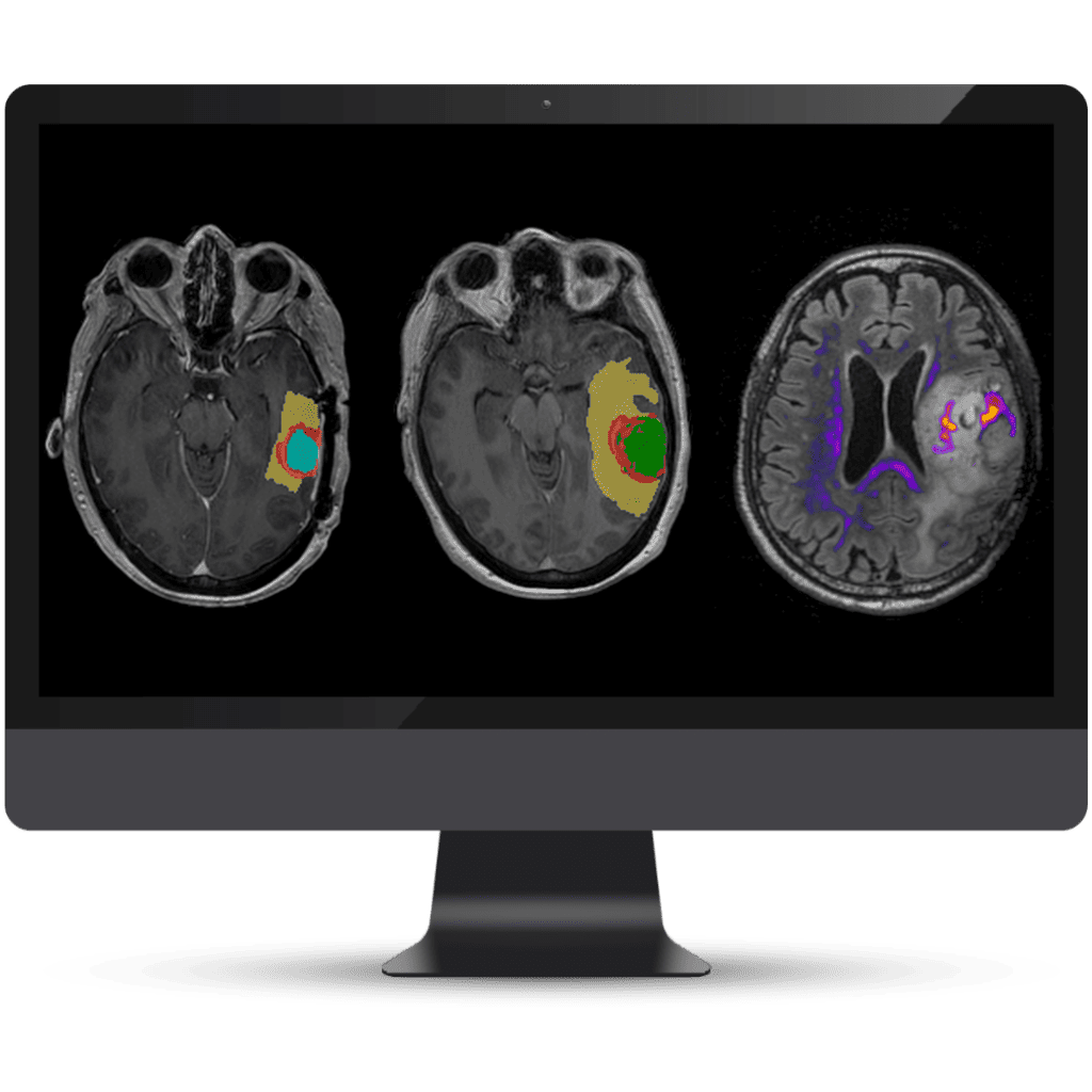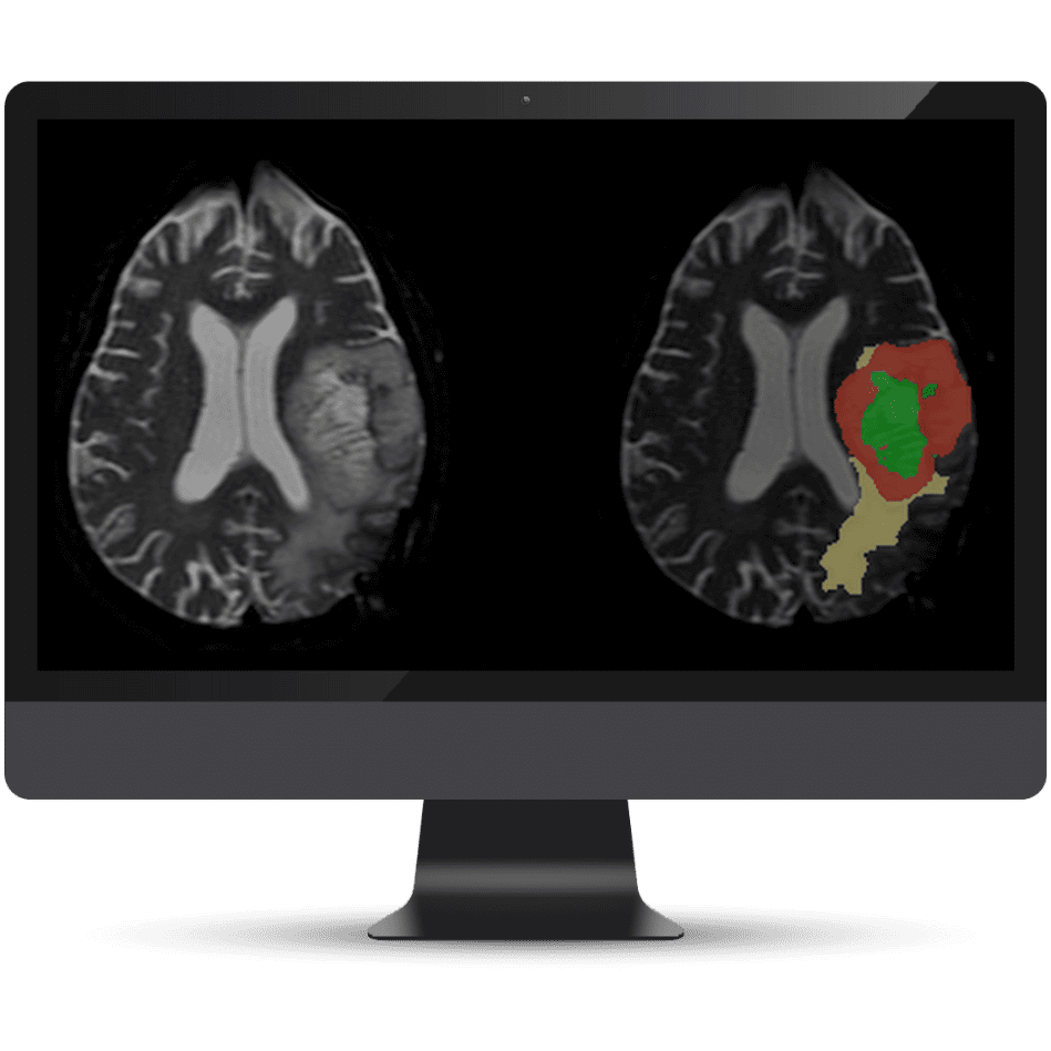
Powered by AI and advanced diffusion MRI, NeuroQuant® Brain Tumor assists radiologists, oncologists, and the Tumor Board by providing objective quantification and analysis of tumor changes over time.
NeuroQuant® Brain Tumor includes tumor segmentations that are color-coded slice by slice and routed to PACS, making it easy for radiologists to identify regions of interest and track changes over time. The integration of Restriction Spectrum Imaging (RSI) provides deeper insights into tumor microstructure, enhancing the analysis with more detailed information than traditional imaging methods.


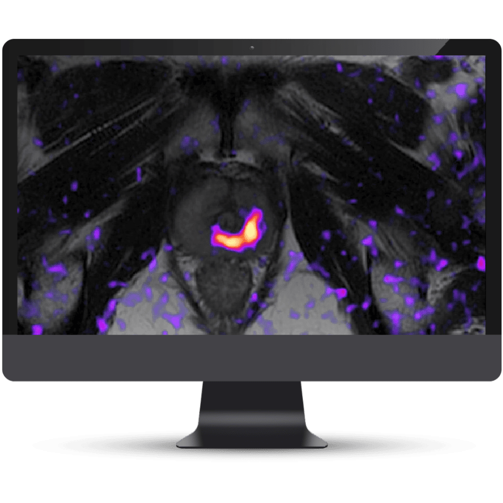
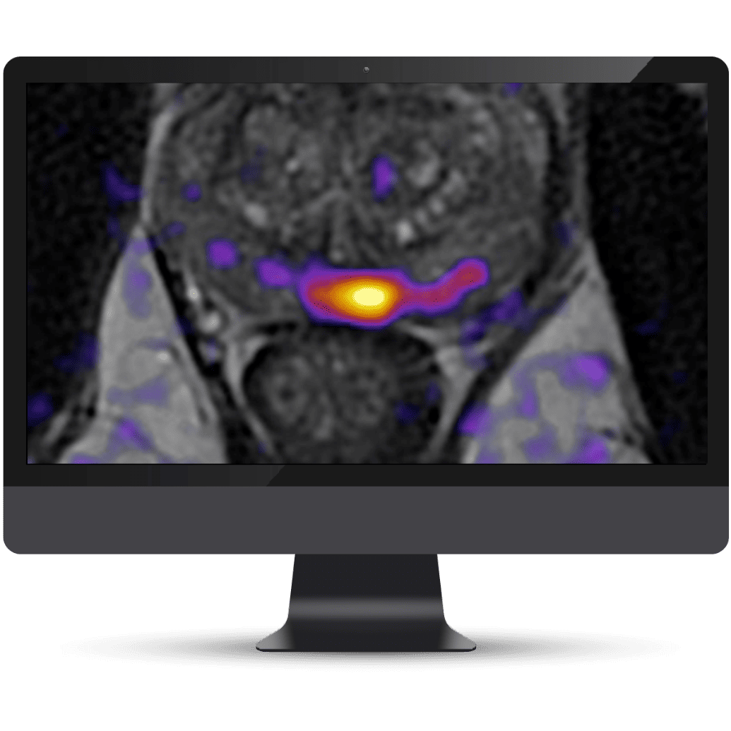
OnQ Prostate is an FDA-cleared software solution using Restriction Spectrum Imaging (RSI), a patented diffusion MRI method that has been shown to improve detection, in vivo characterization, and localization of clinically significant cancer.
Through clear, easy-to-interpret images OnQ™ Prostate empowers collaborative decision-making and enhances targeting for biopsy and therapy. Elevating clinical workflows that use MRI for prostate cancer detection, monitoring, and treatment, it helps bridge the gaps between radiologists and referring physicians.

Use high-resolution 3D T1 images to detect subtle changes in ventricle expansion, tissue atrophy, and swelling.
Monitor structure volumes and visually evaluate changes using color-coded overlays and change quantification data.
Compare the patient’s brain structure volume measurements to a normal population.
Analyze ages 3-100 using our Dynamic Atlas™ technology, validated over thousands of clinical cases.
Tailor solutions for a broad spectrum of neurological disorders, including white matter hyperintensities for Alzheimer’s disease.
Identify confluent lesions and new lesion load
Longitudinal reporting monitors structure volumes and visual changes from previous scans – all within a single report.
Color-coded overlay demonstrates lesion dynamics.
Compare the patient’s brain structure volume measurements to a normal population.
Analyse ages 3-100 using our Dynamic Atlas™ technology, validated over thousands of clinical cases.
©2024 Kryptonite SolutionsTM. All Rights Reserved.
Powered by: Purple Tuché